
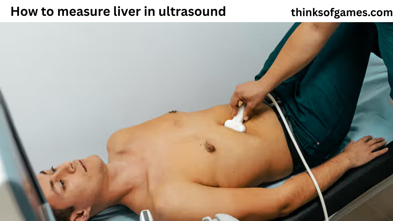
If you are wondering how to measure liver in ultrasound, the process involves much more than placing a measurement line on an ultrasound image. The liver is a large organ with an irregular shape, and its apparent size can change depending on the imaging plane, patient position, breathing, body shape, and technique used by the sonographer.
Ultrasound is commonly used to examine the liver because it is noninvasive, does not use ionizing radiation, and can provide information about liver size, appearance, blood vessels, bile ducts, and focal abnormalities. The American College of Radiology recommends that liver ultrasound examinations include appropriate longitudinal and transverse views and notes that liver measurement may be performed on longitudinal images at the midclavicular line.
Understanding how to measure liver in ultrasound is especially important for sonographers, radiologists, physicians, and medical students. For patients, understanding the basic idea can also make an ultrasound report easier to understand.
A liver measurement is only one part of a complete ultrasound examination. A liver that measures within a commonly used range is not automatically healthy, and a liver that measures larger than expected does not automatically mean that a person has liver disease.
The measurement must always be interpreted together with the patient’s history, laboratory results, symptoms, liver appearance, and other ultrasound findings.
What Is Liver Measurement on Ultrasound?
Liver measurement on ultrasound is an estimate of the size of the liver obtained from an appropriate imaging plane.
Because the liver has a three-dimensional shape, one measurement cannot describe its entire volume.
In routine abdominal ultrasound, the sonographer may obtain a longitudinal image and measure the liver along a standardized anatomical direction. The ACR practice parameter states that liver measurement may be performed on longitudinal images at the midclavicular line.
The exact measurement method can vary between institutions and clinical situations.
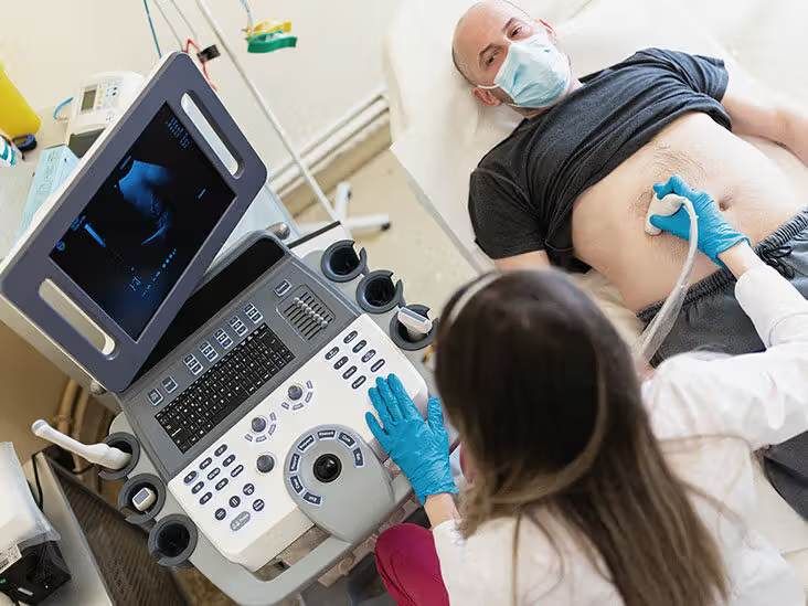
This is important because different techniques can produce different numbers.
For example, a measurement taken in one oblique plane may be longer than a measurement obtained in a more standardized plane. If the patient is breathing differently or the liver is not imaged in the same orientation, the reported number may also change.
Also read:What Causes Fatty Liver Disease? Causes, Risk Factors, Symptoms, and Prevention
Therefore, when learning how to measure liver in ultrasound, technique and consistency are just as important as the final number.
Why Is Liver Size Measured on Ultrasound?
Liver size can provide useful information about the patient’s condition.
A liver may appear enlarged in conditions such as:
- Fatty liver disease
- Hepatitis
- Congestion
- Certain infiltrative diseases
- Some tumors
- Other liver or systemic conditions
A liver may also become smaller or develop an irregular contour in advanced chronic liver disease.
However, liver size alone cannot determine the cause of an abnormality.
The sonographer and radiologist also evaluate the liver’s echotexture, surface contour, blood vessels, bile ducts, focal lesions, and other features.
The ACR abdominal ultrasound practice parameter recommends evaluation of the liver parenchyma for focal and diffuse abnormalities and assessment of major hepatic and perihepatic vessels.
How to Measure Liver in Ultrasound Step by Step?
The exact scanning protocol depends on the examination and the facility, but a general approach can be understood in several stages.
Prepare the Patient
Patient preparation can affect image quality.
For many liver ultrasound examinations, fasting is used to reduce bowel gas and improve visualization of the upper abdomen. ACR guidance for liver ultrasound describes fasting before the examination, while specific requirements can vary by the type of ultrasound and patient situation.
The patient is commonly positioned supine, although additional positions may be used to improve the acoustic window.
The right arm may be raised to increase the space between the ribs during intercostal scanning.
Choose the Appropriate Transducer
A curvilinear abdominal transducer is commonly used for routine liver imaging.
The selected frequency depends on the patient’s body habitus and the depth of the liver.
Higher frequencies can provide greater detail but have less penetration.
Lower frequencies generally provide deeper penetration and may be useful in patients with a larger body habitus.
The sonographer adjusts the equipment according to the patient’s anatomy and image quality.
Obtain a Longitudinal View
A longitudinal view is important when assessing liver size.
The goal is to obtain a representative image of the liver along its long axis rather than measuring an incomplete or artificially elongated portion.
The ACR specifically notes that liver measurement may be performed on longitudinal images at the midclavicular line.
The midclavicular line is an anatomical reference that helps standardize where the measurement is obtained.
Identify the Liver Margins
Before placing calipers, the sonographer needs to identify the relevant superior and inferior margins of the liver.
The measurement should represent the liver dimension being assessed.
Poor visualization of the diaphragm, an incomplete liver edge, or an oblique imaging plane can make the measurement less reliable.
Place the Measurement Calipers
The calipers are positioned from the appropriate superior margin to the appropriate inferior margin along the selected measurement axis.
The measurement is usually recorded in centimeters.
The exact caliper placement and axis should follow the protocol used by the imaging department or supervising radiologist.
The important principle is consistency.
A measurement should not be taken simply along whichever direction produces the largest number.
Record the Measurement
The resulting value is documented in the ultrasound report.
Some reporting systems include liver length as an optional report element, while the overall examination also describes liver appearance and other findings. The ACR’s LI-RADS reporting guidance includes liver length as a report field.
What Is the Midclavicular Line?
The midclavicular line is an imaginary vertical anatomical line that passes downward from approximately the midpoint of the clavicle.
It provides a reproducible reference for certain abdominal measurements.
When learning how to measure liver in ultrasound, the midclavicular line is important because measurements obtained from different locations can vary.
Using a consistent reference point makes comparisons more meaningful.
This becomes especially important when a patient has follow-up ultrasound examinations.
If two examinations use different techniques, a small difference in measured liver length may reflect technique rather than a true change in liver size.
What Is a Normal Liver Size on Ultrasound?
There is no single number that defines a normal liver size for every adult.
Liver size can vary with:
- Height
- Sex
- Body size
- Age
- Body habitus
- Measurement technique
- Imaging plane
- Breathing
- Individual anatomy
A commonly cited clinical reference is around 15 to 16 cm for certain right-lobe craniocaudal measurements, but this should not be treated as a universal cutoff.
The reason is simple: different studies and institutions use different measurement methods.
A number cannot be interpreted properly without knowing how and where it was measured.
This is one of the most important lessons when learning how to measure liver in ultrasound.
Why Can Liver Measurements Differ Between Ultrasounds?
It is normal for measurements to vary somewhat between examinations.
Several factors can contribute.
Different Imaging Planes
A slightly oblique plane can make the liver appear longer.
Different Patient Positions
Changing from supine to another position can change the liver’s relationship with surrounding structures.
Breathing
The liver moves with respiration.
A deep breath may move the liver downward and alter the apparent measurement.
Body Habitus
Excess body tissue or limited acoustic windows can make it difficult to obtain a complete image.
Operator Technique
Different sonographers may select slightly different landmarks or imaging planes.
Equipment Settings
Ultrasound frequency, depth, focus, and other settings can affect visualization.
The ACR recognizes factors such as large body habitus, limited acoustic windows, heterogeneous liver tissue, and reduced beam penetration as reasons liver visualization may be incomplete.
How Important Is the Measurement Technique?
Technique is extremely important.
A technically poor measurement can be misleading even if the number looks precise.
For example, an ultrasound machine may display a measurement such as 16.2 cm, but the decimal point does not mean that the measurement is accurate to a tenth of a centimeter in every situation.
The quality of the image and the correctness of the measurement plane matter first.
This is why trained professionals evaluate the entire image rather than relying on a single number.
Should the Liver Be Measured in One or More Directions?
Routine liver assessment includes multiple imaging planes.
The ACR practice parameter recommends long-axis and transverse views of the liver.
However, measuring liver length is different from calculating total liver volume.
A single length measurement does not represent the complete three-dimensional volume of the organ.
If an accurate liver volume is clinically needed, other imaging methods or specialized volumetric techniques may be used.
The ACR has described hepatic volume assessment as an area where computer-assisted segmentation can potentially improve reproducibility, particularly in settings such as surgical planning and chronic liver disease evaluation.
How to Measure Liver in Ultrasound for the Right Lobe?
The right hepatic lobe is often the focus of routine liver length measurement because it provides a relatively accessible acoustic window.
A longitudinal image is obtained through an appropriate anatomical reference, commonly around the midclavicular line.
The sonographer identifies the superior and inferior extent of the liver and places the measurement along the appropriate long axis.
The exact technique should follow the department’s protocol.
This is important because there is no value in memorizing one measurement technique while ignoring the imaging protocol used by the interpreting radiologist.
Can the Left Lobe Be Measured Too?
Yes, the left lobe can be assessed and measured when clinically appropriate.
However, left-lobe measurements are not necessarily interchangeable with the standard liver length reported in routine abdominal ultrasound.
The left lobe has a different shape and orientation.
A left-lobe dimension therefore needs to be labeled clearly so that it is not confused with the overall liver length or right-lobe measurement.
A complete liver ultrasound should evaluate both the right and left lobes rather than focusing only on one measurement.
Current ACR accreditation requirements emphasize complete longitudinal and transverse documentation of the liver, including multiple views of the right and left lobes.
How to Measure Liver in Ultrasound When the Patient Is Obese?
Patients with larger body habitus can present a technical challenge.
Ultrasound waves must travel farther through tissue, and image quality may decrease.
The sonographer may need to:
- Adjust transducer frequency
- Change patient position
- Ask the patient to change breathing
- Use an intercostal window
- Apply appropriate probe pressure
- Adjust imaging settings
- Try different acoustic windows
ACR guidance specifically recommends adjusting patient positioning, inspiration level, acoustic window, probe pressure, and ultrasound settings when liver visualization is difficult.
A limited examination should be recognized as limited rather than forcing an unreliable measurement.
How to Measure Liver in Ultrasound With Poor Visualization?
Sometimes the entire liver cannot be clearly seen.
Reasons can include:
- Bowel gas
- Obesity
- Limited patient cooperation
- Rib shadowing
- Poor acoustic windows
- Liver heterogeneity
- Reduced penetration
The correct response is not simply to guess the liver margin.
The sonographer should attempt appropriate technical adjustments.
If the liver remains poorly visualized, the limitation should be documented.
The ACR LI-RADS ultrasound system specifically recognizes visualization limitations and uses visualization scores to describe whether technical limitations may reduce the ability to detect liver observations.
Why Is Patient Position Important?
Patient position can change the acoustic window.
The liver sits primarily in the upper right abdomen beneath the diaphragm and ribs.
When the patient changes position or raises an arm, the intercostal spaces and relationship between the liver and abdominal structures may change.
This can make certain parts of the liver easier to see.
For liver elastography, current ACR guidance describes supine or slight lateral positioning with the right arm raised to improve the intercostal space.
Routine liver ultrasound may use additional positioning as needed.
Why Does Breathing Matter During Liver Ultrasound?
Breathing changes the position of the liver.
During inspiration, the diaphragm moves downward, and the liver can move with it.
This movement may create a better acoustic window.
A sonographer may ask the patient to take a deep breath and hold it briefly.
The goal is to place the liver in a position that allows better visualization.
However, the patient should only hold their breath as instructed and should not be uncomfortable.
How Does Liver Shape Affect Measurement?
The liver is not a simple rectangular or oval organ.
Its shape is complex, with different lobes and segments.
This is why measuring it with a simple straight line does not tell the entire story.
A liver can have a relatively normal length but still show abnormal contour, altered echotexture, or focal abnormalities.
Likewise, a mildly increased measurement does not automatically mean that the liver is diseased.
The measurement is only one part of the examination.
What Else Is Checked During a Liver Ultrasound?
A complete liver ultrasound evaluates much more than size.
The sonographer and radiologist may assess:
- Liver echogenicity
- Liver texture
- Surface contour
- Focal lesions
- Hepatic veins
- Portal vein
- Inferior vena cava
- Bile ducts
- Gallbladder
- Adjacent structures
- Ascites
The ACR practice parameter recommends evaluation of the major hepatic and perihepatic vessels, hepatic lobes, and other relevant structures.
This broader assessment is why a liver ultrasound report should never be interpreted from the liver measurement alone.
What Does Liver Echogenicity Mean?
Echogenicity describes how bright the liver appears on ultrasound.
The liver is commonly compared with the right kidney when assessing echogenicity.
The ACR practice parameter specifically recommends comparing liver parenchymal echogenicity with the right renal cortex when possible.
Increased liver echogenicity can be associated with fatty infiltration, although ultrasound appearance is not perfectly specific.
The ACR notes that conventional ultrasound can detect moderate and severe steatosis reasonably well but is less reliable for mild steatosis, and interpretation can be affected by inflammation and fibrosis.
This is another example of why a single ultrasound finding should not be interpreted in isolation.
Can Ultrasound Diagnose Fatty Liver?
Ultrasound can provide evidence suggesting fatty liver, especially when fat accumulation is more than mild.
However, conventional ultrasound has limitations.
The ACR notes that ultrasound can be unreliable for mild fatty infiltration and that sensitivity and specificity decrease when mild disease is included.
A report describing increased liver echogenicity does not automatically determine the amount of fat in the liver.
Depending on the clinical situation, additional evaluation may be appropriate.
Can Liver Size Diagnose Fatty Liver?
No.
A liver may be enlarged in some people with fatty liver, but size alone cannot diagnose fatty liver.
A person can have fatty liver without a dramatically enlarged liver.

Similarly, an enlarged liver can have causes other than fat accumulation.
The diagnosis requires consideration of the ultrasound appearance, medical history, laboratory findings, and other clinical information.
How to Measure Liver in Ultrasound for Follow-Up Exams?
Consistency is especially important during follow-up.
Also read:How to Stop Liver Pain Immediately at Home: Safe Steps, Causes, and When to Get Help
If a patient has an ultrasound today and another one months later, the two measurements are easier to compare when similar techniques are used.
The sonographer should use an appropriate and reproducible measurement plane.
The interpreting physician can then consider whether an apparent change is likely to represent a true change.
ACR guidance emphasizes standardized protocols and comparison with prior examinations when possible for liver ultrasound assessment.
Can Liver Size Change Over Time?
Yes.
The liver can change in size because of disease, treatment, body changes, or other factors.
But small differences between ultrasound examinations do not always mean the liver has truly changed.
Measurement variation is common.
For example, a difference of several millimeters may be related to breathing or plane selection rather than biological growth or shrinkage.
This is why trends should be interpreted by a radiologist or physician rather than by comparing numbers without context.
What Does an Enlarged Liver Mean?
An enlarged liver is called hepatomegaly.
Hepatomegaly is a finding, not a diagnosis.
Possible causes include:
- Fat accumulation
- Inflammation
- Congestion
- Certain infections
- Infiltrative diseases
- Some tumors
- Other medical conditions
If an ultrasound report says the liver is enlarged, the next step is to understand why.
The doctor may consider blood tests, medical history, medications, symptoms, and other imaging findings.
What Does a Small Liver Mean?
A small or shrunken liver can sometimes be seen in advanced chronic liver disease, especially when accompanied by other abnormal features.
However, a measurement that appears small does not automatically mean cirrhosis.
Again, technique and patient anatomy matter.
The liver’s contour and internal appearance can be more informative than size alone in certain conditions.
What Is Liver Surface Nodularity?
The liver surface can be evaluated for smoothness or nodularity.
An irregular or nodular liver contour can be associated with chronic liver disease and cirrhosis.
The ACR practice parameter notes that the liver surface may be examined with a high-frequency transducer when evaluating patients at risk for cirrhosis.
This illustrates another important point: liver ultrasound is not simply about measuring centimeters.
The shape and surface of the liver also matter.
How Are Liver Lesions Measured on Ultrasound?
A focal liver lesion is measured differently from the overall liver length.
If a lesion is found, its size is generally documented in multiple dimensions.
The ACR LI-RADS ultrasound recommendations state that focal liver observations should be documented in three dimensions and that the involved lobe and segment should be recorded when possible.
The measurement should be taken where the lesion margins are best visualized.
For follow-up, using a consistent measurement approach can help evaluate whether the lesion has changed.
Why Is the Portal Vein Important?
The portal vein is one of the major blood vessels supplying the liver.
Ultrasound can evaluate its patency, direction of flow, and other characteristics.
Color Doppler and spectral Doppler may be used when vascular assessment is indicated.
The ACR abdominal ultrasound practice parameter recommends Doppler evaluation for vascular examinations, including assessment of portal and hepatic vessels.
This information can be important in patients with suspected or known liver disease.
How Is Liver Stiffness Different From Liver Size?
Liver size and liver stiffness are two completely different measurements.
Liver size describes the physical dimensions of the organ.
Liver stiffness evaluates the mechanical properties of liver tissue.
Elastography is an ultrasound-based technique that can be used to assess liver stiffness.
Stiffness measurements may help evaluate fibrosis, depending on the clinical setting.
A patient can have a normal liver size but increased stiffness.
Likewise, an enlarged liver does not automatically mean that stiffness is increased.
What Is Ultrasound Elastography?
Ultrasound elastography estimates tissue stiffness.
Different elastography methods exist, including point shear-wave elastography and two-dimensional shear-wave elastography.
Current ACR guidance recommends appropriate technique, patient positioning, and measurement location for liver stiffness examinations.
Elastography is not the same as measuring liver length.
The two measurements answer different clinical questions.
Why Is the Liver Measured in Centimeters?
Centimeters provide a simple numerical description of liver length.
A standardized unit makes it easier to communicate findings between healthcare professionals and compare examinations.
However, a measurement such as 15.4 cm should not be interpreted without knowing:
- Where it was measured
- Which imaging plane was used
- Patient size
- Patient age
- Clinical situation
- Other ultrasound findings
The number is useful because it is standardized, but the context gives it meaning.
What Should a Liver Ultrasound Report Include?
A liver ultrasound report may describe:
- Liver size
- Liver echogenicity
- Liver texture
- Surface contour
- Focal abnormalities
- Portal vein
- Hepatic vessels
- Bile ducts
- Gallbladder
- Ascites
- Technical limitations
The exact report depends on the type of examination.
For liver cancer surveillance, ACR LI-RADS provides standardized terminology and reporting systems designed to improve consistency.
What Does a Limited Liver Ultrasound Mean?
A limited study means that technical factors prevented complete or ideal visualization.
It does not necessarily mean that the patient has more severe liver disease.
For example, bowel gas or body habitus may prevent the entire liver from being seen clearly.
The ACR LI-RADS system specifically recognizes moderate and severe visualization limitations because they can reduce the ability of ultrasound to detect small liver observations.
If an examination is limited, the physician may recommend another imaging test depending on the clinical question.
How to Measure Liver in Ultrasound Correctly?
The safest summary is to follow a standardized imaging protocol rather than relying on a single memorized number.
A trained professional should:
- Prepare and position the patient appropriately.
- Select a suitable transducer.
- Obtain representative longitudinal and transverse liver images.
- Use an appropriate anatomical reference for liver length.
- Obtain the measurement on a suitable longitudinal image.
- Place calipers along the appropriate liver axis.
- Record the value in centimeters.
- Evaluate the rest of the liver and upper abdomen.
- Document technical limitations.
- Interpret the measurement together with the complete clinical and imaging picture.
The ACR’s current abdominal ultrasound guidance supports longitudinal and transverse liver imaging and allows liver measurement on longitudinal images at the midclavicular line.
Common Mistakes When Measuring the Liver
Measuring an Oblique Plane
An oblique plane can produce a misleadingly large measurement.
Also read:Recipes for Healthy Liver: Easy, Nutritious Meals for Better Liver Health
Measuring the Wrong Structure
The calipers must be placed on the actual liver margins.
Ignoring Patient Position
Position can affect the acoustic window and apparent anatomy.
Ignoring Breathing
Respiratory motion can change liver position.
Using One Number as a Diagnosis
Liver length does not diagnose liver disease by itself.
Comparing Different Techniques
Measurements from different protocols may not be directly comparable.
Ignoring Image Quality
A precise-looking number from a poor-quality image is not necessarily reliable.
Can Patients Measure Their Own Liver With an Ultrasound Machine?
Patients should not attempt to diagnose or measure their own liver using consumer ultrasound equipment.
Professional liver ultrasound requires knowledge of anatomy, scanning technique, image optimization, and interpretation.
Even obtaining a number is not enough.
The person interpreting the examination must understand whether the measurement was technically valid and what other findings mean.
If you have received a liver measurement on an ultrasound report, the safest approach is to discuss it with your healthcare professional.
Does Liver Measurement Tell You If Your Liver Is Healthy?
No.
Liver health cannot be determined from size alone.
A complete assessment may involve:
- Symptoms
- Medical history
- Blood tests
- Ultrasound findings
- Elastography
- Other imaging
- Risk factors
- Physical examination
- Sometimes additional testing
A normal liver measurement does not guarantee normal liver function.
Likewise, a slightly enlarged liver does not automatically indicate serious disease.
What Should You Do If Your Ultrasound Says Your Liver Is Enlarged?
Do not panic.
An enlarged liver can have many causes, and the finding needs to be interpreted in context.
Ask your healthcare professional:
- How enlarged is the liver?
- How was it measured?
- Does the liver have fatty changes?
- Is the surface smooth?
- Are there focal lesions?
- Are the portal and hepatic vessels normal?
- Are my liver blood tests normal?
- Do I need additional testing?
These questions are more useful than focusing only on the number.
How to Read a Liver Measurement on an Ultrasound Report
Suppose a report states that the liver measures 16 cm.
That number should not be interpreted alone.
Look at the rest of the report.
Does it mention:
- Normal echogenicity?
- Increased echogenicity?
- Fatty infiltration?
- Smooth contour?
- Nodular contour?
- Focal lesions?
- Normal portal vein?
- Normal bile ducts?
- Technical limitations?
The overall impression is generally more useful than one isolated measurement.
Why Standardization Matters in Liver Ultrasound
Standardization makes medical imaging more reproducible.
If every sonographer used a completely different method, comparing results would be difficult.
Professional organizations such as the American College of Radiology publish practice parameters to reduce unnecessary variation and support consistent imaging quality.
The ACR’s current ultrasound accreditation requirements also emphasize complete documentation of the liver in longitudinal and transverse planes.
Standardization is especially important when imaging is used for follow-up.
How to Measure Liver in Ultrasound for Students
Students learning abdominal ultrasound should understand the liver anatomy before memorizing measurement numbers. A simple learning process is to first study normal liver anatomy and then learn how the liver looks in longitudinal and transverse views. Next, learn to identify the diaphragm and liver edges, understand the midclavicular reference, and practice obtaining a proper long-axis image. After that, focus on correct caliper placement and compare measurements from different views. Finally, learn to consider liver size along with its echogenicity, contour, blood vessels, and any focal findings. This is more useful than simply memorizing one number as “normal.”
What Is the Difference Between Liver Length and Liver Volume?
Liver length is a one-dimensional measurement.
Liver volume is a three-dimensional estimate.
A liver is not a simple geometric object, so calculating its exact volume from one length is not reliable.
When liver volume is clinically important, specialized imaging and volumetric techniques may be used.
The ACR has highlighted hepatic volume assessment as an area where automated or computer-assisted segmentation may improve reproducibility and efficiency.
Why Ultrasound Is Useful for Liver Examination
Ultrasound has several advantages.
It is:
- Noninvasive
- Widely available
- Generally less expensive than many advanced imaging tests
- Free of ionizing radiation
- Useful for real-time imaging
- Capable of Doppler assessment
It can provide information about liver structure and blood flow.
However, ultrasound also has limitations.
Image quality can be reduced by body habitus, bowel gas, rib shadows, and other technical factors.
That is why ultrasound findings sometimes need to be supplemented by CT, MRI, laboratory testing, or other evaluation.
What Are the Limitations of Measuring Liver Size?
The biggest limitation is that the liver is a complex three-dimensional organ.
A single length does not represent its entire size.
Measurement also depends on technique.
Different operators may obtain somewhat different values.
Patient anatomy can also affect the measurement.
For these reasons, liver size should be treated as one imaging feature rather than an absolute measure of liver health.
Final Thoughts
Learning how to measure liver in ultrasound starts with understanding that the measurement is only one part of a complete liver examination. In routine abdominal ultrasound, the liver is evaluated in longitudinal and transverse planes, and liver length may be measured on a suitable longitudinal image using an anatomical reference such as the midclavicular line.
A reliable measurement depends on proper patient positioning, a good acoustic window, appropriate equipment settings, correct identification of the liver margins, and consistent caliper placement. Current ACR guidance also emphasizes complete liver documentation and recognition of technical limitations that can affect visualization.
Most importantly, a liver measurement should never be interpreted by itself. Liver appearance, echogenicity, surface contour, blood vessels, bile ducts, focal findings, symptoms, laboratory results, and medical history all help determine what the ultrasound means.
For patients, the best approach is to discuss an abnormal liver measurement with the healthcare professional who ordered or interpreted the examination. For students and imaging professionals, following a standardized institutional protocol and current professional guidance is essential for producing reliable and reproducible measurements.
Frequently Asked Questions About How to Measure Liver in Ultrasound
Where is liver length usually measured on ultrasound?
Liver length may be measured on a longitudinal image at the midclavicular line. The exact technique should follow the imaging department’s protocol and the interpreting physician’s requirements.
Is a 15 cm liver normal?
A liver measurement around 15 cm can fall within commonly used reference ranges for certain measurement techniques, but there is no single universal cutoff for every adult. Patient size, sex, age, anatomy, and measurement technique all matter.
Can ultrasound tell if the liver is fatty?
Ultrasound can show findings that suggest fatty liver, particularly when fat accumulation is more than mild. However, conventional ultrasound is less reliable for mild steatosis and cannot determine the exact amount of liver fat by size measurement alone.
Can liver size change between two ultrasound examinations?
Yes. True changes can occur, but small differences can also result from different operators, breathing, patient position, imaging planes, or technical factors. Comparing examinations is most useful when similar techniques are used.
Is liver measurement the same as liver stiffness measurement?
No. Liver measurement describes the physical size of the organ, while liver stiffness is assessed with techniques such as ultrasound elastography. They provide different types of information and should not be confused.

Key Takeaways Reliable financial AI depends on accurate, traceable, well-permissioned [...]

Key Takeaways Put safety and medical care ahead of evidence [...]

Key Takeaways Living alone can be safe when daily routines, [...]
Atlanta patients often receive care from large hospital systems, neighborhood [...]
Diagnostic Precision and Risk Stratification in Modern Urology The foundation [...]

Key Takeaways A recurring SUV problem should be documented from [...]

A printed guide succeeds when it helps someone complete a [...]

Key Takeaways An arrest or citation is an allegation, not [...]